











Clinical–based design of additively manufactured custom sports mouthguards


Dr. Theodoros Tasopoulos
Dr. Olga Naka
Introduction
Sports-related dental injuries represent a substantial public health concern, with studies indicating that up to one-third of oral and craniofacial trauma is associated with athletic activity [1]. Sports mouthguards play a crucial role in preventive dentistry by dispersing and absorbing impact forces, thereby protecting dentofacial and
craniofacial structures [2,3]. Sports mouthguards are generally classified into three main types: stock, mouth-formed (also known as boil-and-bite), and custom-made [4–6]. Custom-made mouthguards are preferred among these categories due to their superior fit, enhanced comfort, and greater protective capabilities, especially in high-impact sports [7]. The European Association for Sports Dentistry (EA4SD) has recently advocated the use of custom-made sports mouthguards rather
than prefabricated or boil-and-bite alternatives, underscoring their efficacy in injury prevention [8]. The efficacy of a mouthguard largely depends on its material composition, thickness, and the manufacturing process employed [9]. Ethylene-vinyl acetate (EVA) has long been the preferred material due to its remarkable impact resistance and inherent flexibility [6,10]. However, EVA mouthguards fabricated by thermoforming frequently exhibit thickness inconsistencies, primarily due to material sagging and model cast distortion during pressure or vacuum forming, particularly in the occlusal areas [11–14]. In response to the constraints posed by conventional manufacturing methodologies, digital technologies have emerged as an alternative for the design and fabrication of sports mouthguards. CAD/CAM workflows, particularly those incorporating additive manufacturing, provide enhanced control over mouthguard thickness and occlusal relationships.
Moreover, these advanced methodologies effectively mitigate the dimensional inaccuracies commonly observed with thermoforming, thereby improving the final product’s accuracy and reproducibility [6,11,15]. This article presents a clinical technique report describing the feasibility and clinical implementation of a fully digital CAD/CAM workflow for the fabrication of custom sports mouthguards. Rather than providing a comparative or outcome-based clinical evaluation, the focus is on detailing each step of the digital process—from intraoral scanning and computer-aided design to additive manufacturing and clinical delivery—highlighting its practical application and reproducibility in a clinical setting. By outlining this workflow, the manuscript aims to offer clinicians a structured, implementable approach to incorporating digital technologies into the fabrication of sports mouthguards.
Materials and methods
A 25-year-old dentate patient engaged in a high-impact contact sport
was presented to our clinic. Clinical examination revealed a stable occlusion,
with no reported parafunctional habits and no signs or symptoms
of temporomandibular disorders. These standardised clinical
conditions allowed for an objective evaluation of the accuracy and
clinical feasibility of the proposed fully CAD/CAM workflow. Written
informed consent was obtained from the patient for the clinical procedures
and for publication of the clinical data and images.
A “New case” was created, and the “Appliance” category was selected in the design software (Splint Studio; 3Shape Unite). Maxillary and mandibular complete-arch intraoral digital scans were acquired using an intraoral scanner (TRIOS 5; 3Shape A/S) according to the manufacturer’s recommended protocol, under optimal ambient lighting conditions.
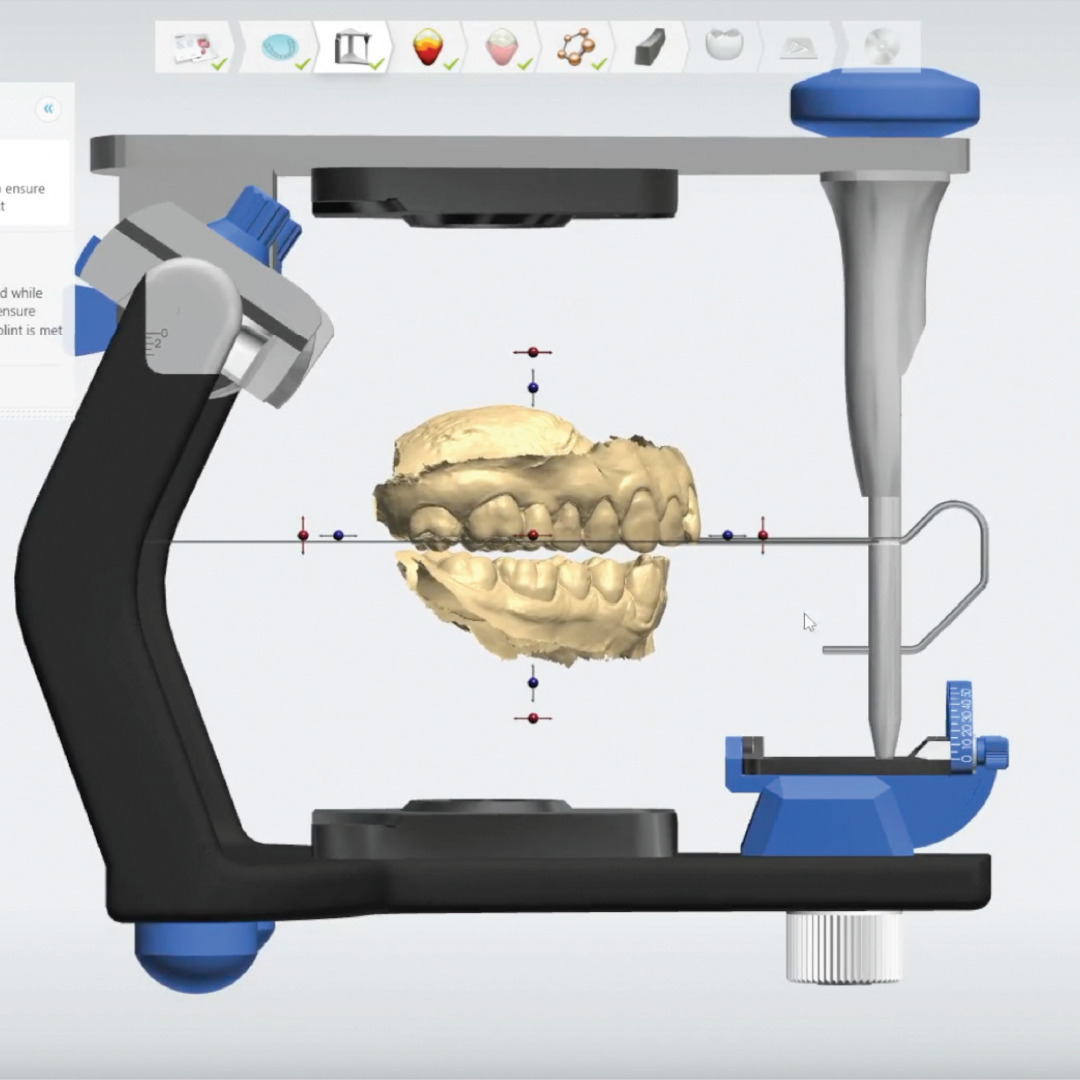
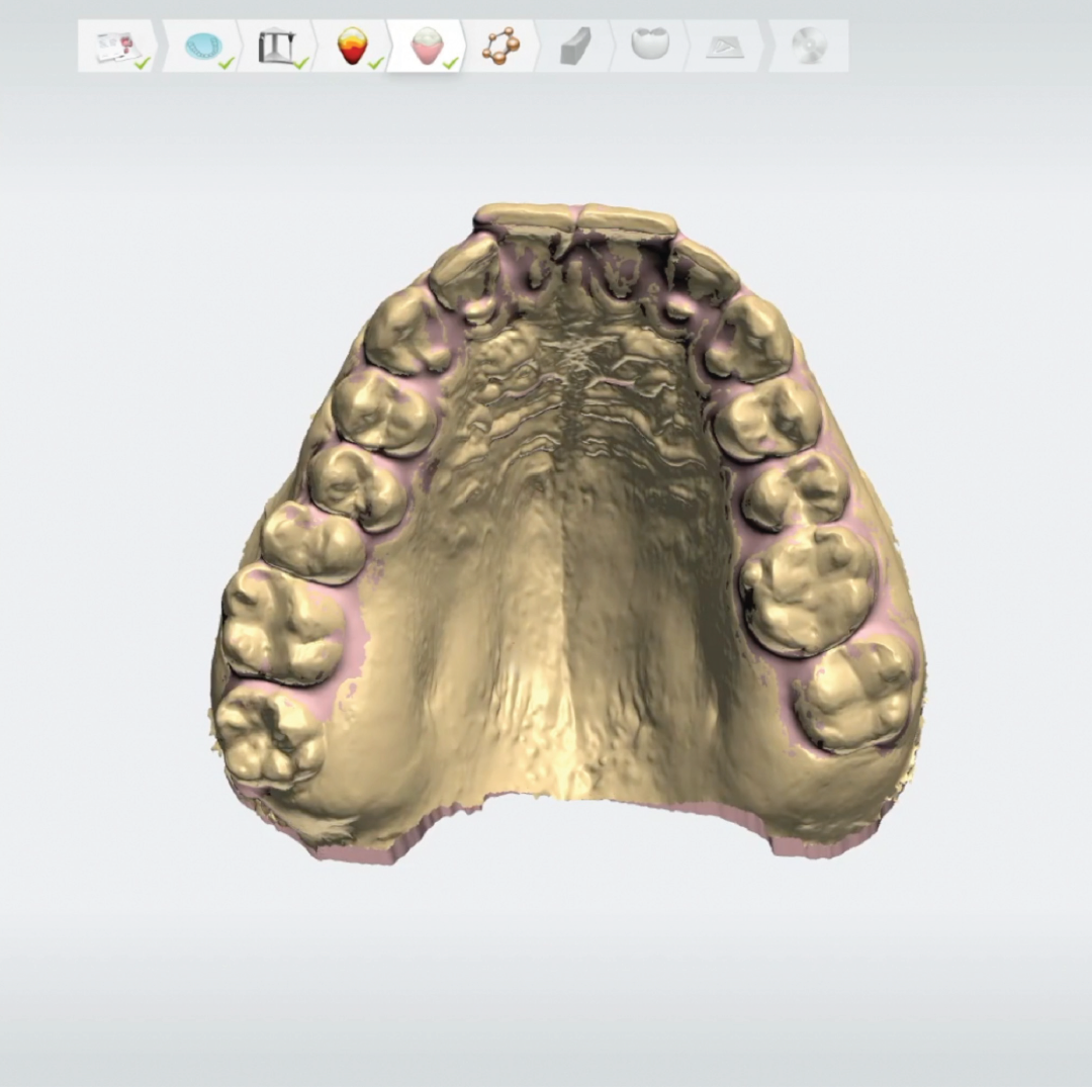
The occlusal vertical dimension (OVD) was increased by 2 mm vertically, using a 3D-printed anterior deprogrammer, thereby establishing. sufficient prosthetic space for the sports mouthguard. A new maxillomandibular relationship was digitally registered at the increased OVD in Centric Relation (CR) using a 3D-printed anterior deprogrammer, designed to encompass at least two maxillary teeth and one mandibular tooth. A vertical raised bilateral occlusal record was acquired, incorporating at least four mandibular and four maxillary teeth. The articulated scans were processed automatically.
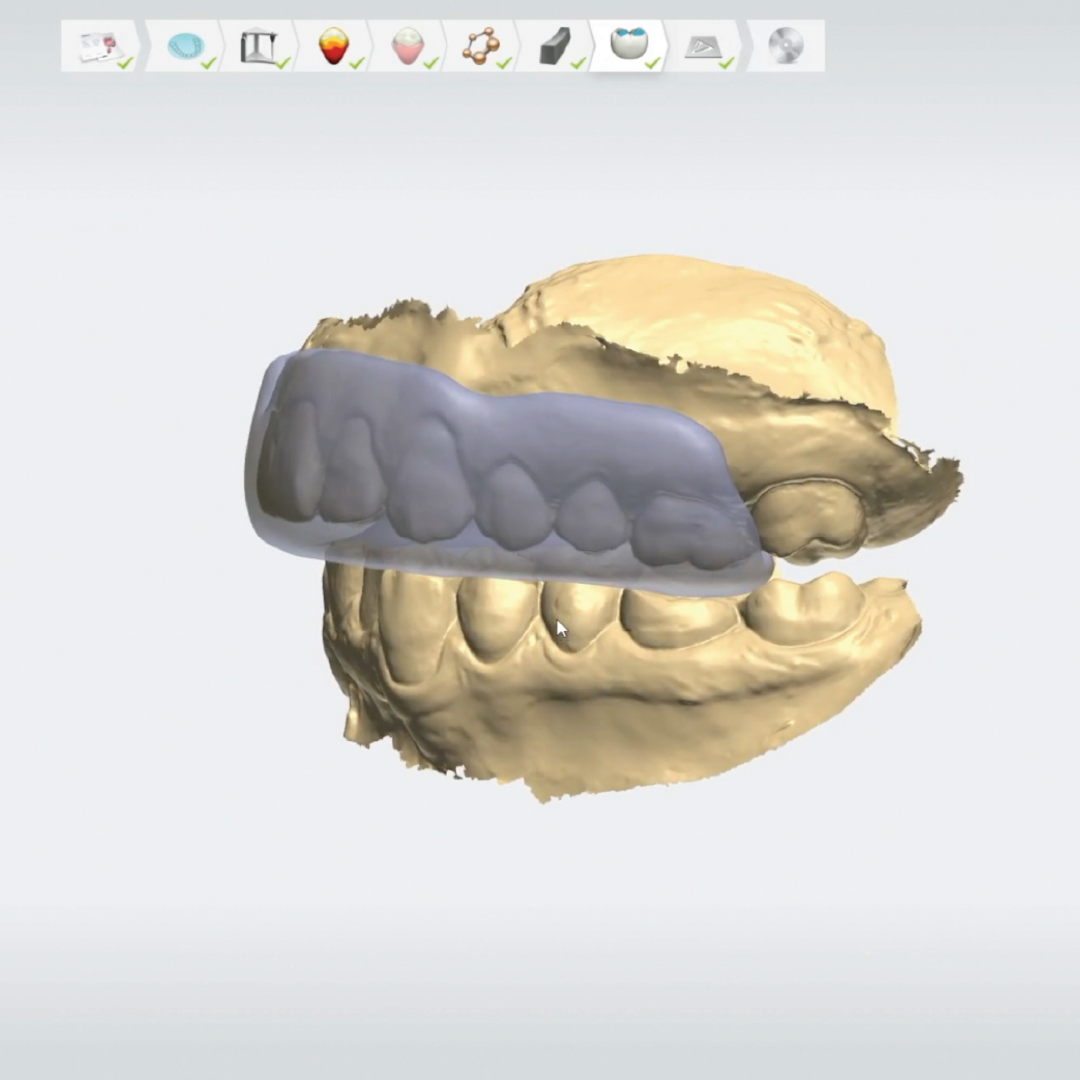
Initially, the occlusal plane was digitally established using the software’s
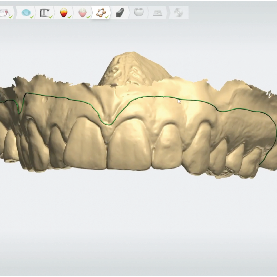
mean – value virtual articulator to articulate the maxillary and mandibular casts in centric relation at the increased occlusal vertical dimension. Subsequently, the insertion path was defined to ensure proper retention and to minimize undercut engagement during placement and removal. Critical interdental spaces were blocked out, and retentive undercuts were reinforced to enhance stability and avoid preventing thin or weak areas within the appliance. The outline of the mouthguard was delineated, and the initial thickness was designated based on the desired protection level. A well-defined retention area was created on the labial surface of the anterior teeth to improve mechanical stability during functional use.
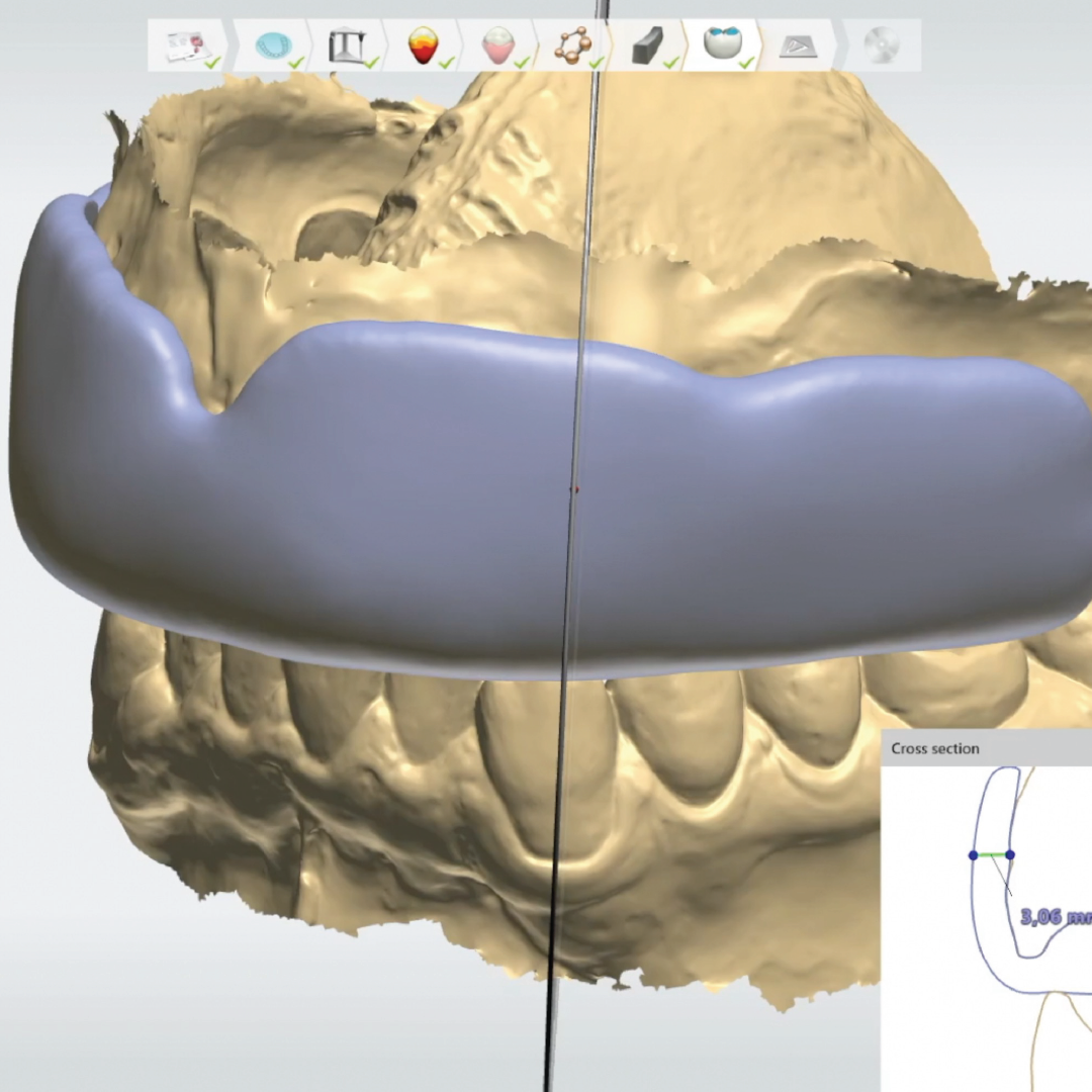
The mouthguard extension was computer-aided designed to cover the distal surfaces of the first molars while maintaining a 2-mm distance from the vestibular sulcus to avoid impingement on the movable mucosa. Labial, occlusal, and palatal thicknesses were set at 3–5 mm, 2 mm, and 1 mm, respectively, in accordance with current recommendations [10–12]. This configuration enhances impact energy absorption at the labial and occlusal surfaces while keeping the palatal aspect thinner to improve comfort and speech. The appliance thickness was then augmented, ensuring that occlusal contacts with the antagonists were appropriately established for the device’s functional integration.
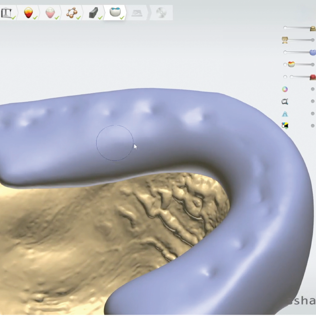
Additionally, selective reduction of the palatal flanges and tapering the appliance margins toward the gingival margins minimises soft-tissue irritation and improves patient comfort. Additionally, selective tapering of the palatal flanges toward the gingival margins minimised soft-tissue irritation and improved patient comfort. Selective reduction of the appliance material in areas corresponding to the palatal cusps of the premolars and molars. Eliminated occlusal-controlled sinking of the mandibular teeth into the appliance, thereby creating a protective barrier for the lower dentition.
A buccal barrier was incorporated to protect the antagonistic dentition
during occlusal loading. Once the design was verified, the complete digital mouthguard file was finalised and exported for manufacturing.
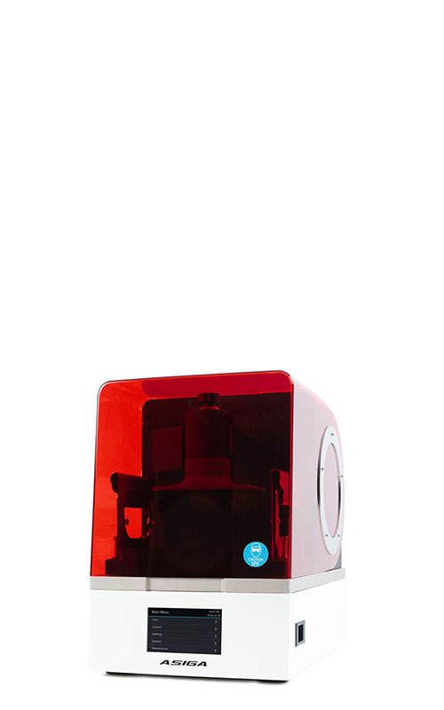
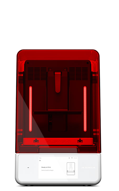
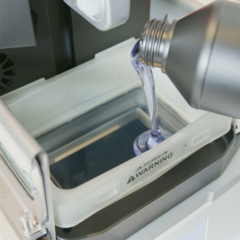
A 3D printer (Asiga Max 2; Asiga) fabricated the mouthguard with a biocompatible resin material specifically designed for this purpose (Keyguard; Keystone Industries). The printing procedure was performed with a 45-degree build angle and a layer thickness of 100 μm. Postprocessing consisted of cleaning in 99% isopropyl alcohol for 5 min, air-drying at room temperature, UV post-curing for 20 min, in accordance with manufacturer-validated parameters. Following printing, all support structures were carefully removed. Finishing and polishing procedures were performed using a Scotch-Brite™ Medium wheel and a Fine Blue Abrasion Disc (Keystone Industries, Gibbstown, NJ, USA).
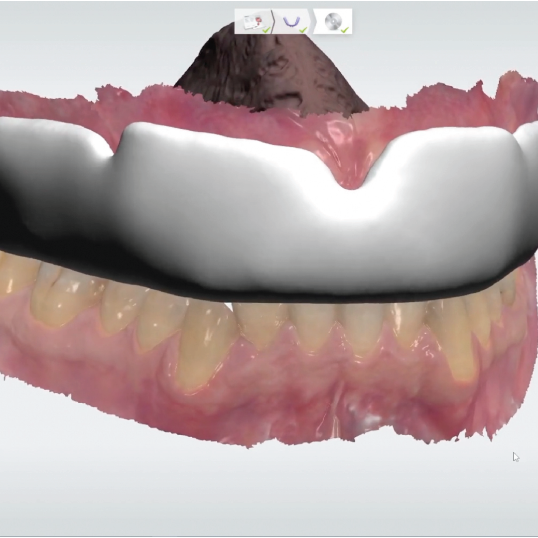
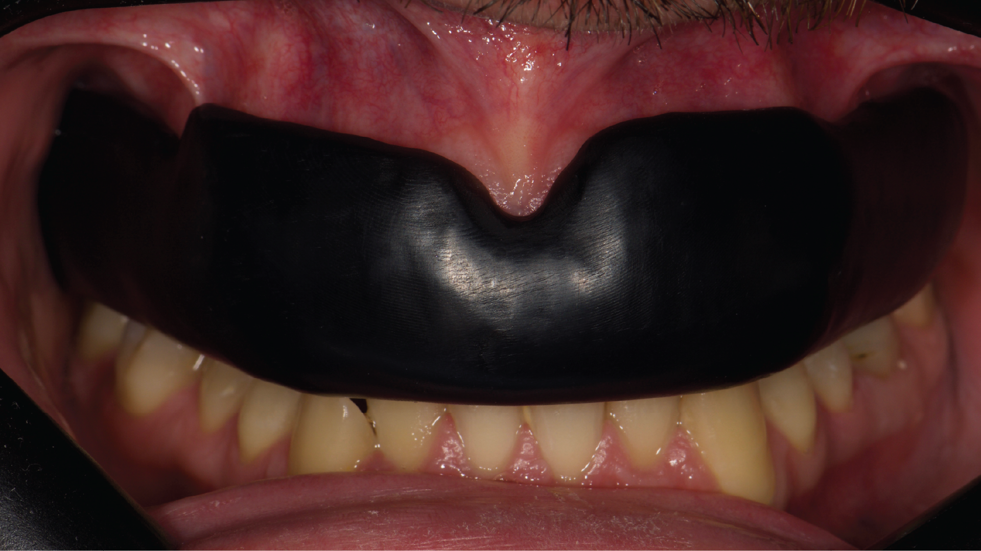
The final delivery of the custom-fitted, additively manufactured sports mouthguard was conducted following a thorough assessment, which included evaluating fit precision, occlusal effectiveness, and comfort for the athlete. No adjustments were required, reflecting the accuracy of the computer-aided design and manufacturing protocol. Fit was assessed through visual inspection of marginal adaptation, stability during insertion and removal, and the absence of localised pressure areas or tissue impingement. Occlusion was evaluated by verifying uniform and stable contacts with the opposing dentition at the increased occlusal vertical dimension and by identifying and eliminating any occlusal interferences.
Results
Patient comfort was assessed based on subjective feedback during initial insertion, functional mandibular movements, speech, and shortterm use during athletic activity. Follow-up appointments were scheduled for 1 month, 6 months, and 1 year. No complications, material degradation, or need for adjustment were observed. This evaluation ensures that the mouthguard not only conforms to the athlete’s specific anatomical needs but also promotes optimal oral health and performance during athletic activities.
Discussion
The implementation of a fully digital CAD/CAM workflow for the fabrication of custom sports mouthguards allows precise control over appliance geometry, thickness distribution, and occlusal relationships, addressing well-documented limitations of conventional thermoforming techniques. Vacuum- or pressure-formed ethylene-vinyl acetate (EVA) mouthguards frequently exhibit thickness variability due to material sagging, uneven pressure distribution, and cast distortion during heating and forming, particularly in the occlusal and incisal regions [12–14]. In contrast, digital workflows enable predefined and uniform thickness values, which have been shown in in vitro studies to improve structural consistency and protective performance [16–18].
The present technique builds upon previously reported digital and semi-digital workflows by integrating intraoral scanning, computer-aided design, and additive manufacturing into a fully in-house clinical protocol. As demonstrated by Ntovas et al. [15], digital occlusal management can improve the functional integration of additively manufactured sports mouthguards, while Rondon et al. [19] reported superior adaptation of digitally fabricated mouthguards to dental surfaces compared with conventional workflows. By using CAD splint software, the proposed approach enables controlled labial, occlusal, and palatal thickness distribution in accordance with current biomechanical recommendations, while allowing precise occlusal contact design at an increased occlusal vertical dimension.
Moreover, the digital storage of the finalized design permits exact reproduction of the appliance without additional clinical or laboratory procedures, enhancing standardisation and efficiency. Compared with laboratory-dependent or partially digital approaches described in the literature [15–21], the workflow presented in this technique article minimises manual variability, reduces chairside adjustments, and supports predictable clinical delivery. These advantages are particularly relevant for athletes who require consistent protection, rapid replacement, and high levels of comfort and retention. Comparative in vitro assessments have shown that 3D-printed mouthguards fit the incisal and lingual surfaces significantly better than those produced on plaster casts [19]. Improved adaptation to the dentition can increase comfort, retention, and overall compliance among athletes. Furthermore, the digital process enables rapid, cost-efficient, and scalable production of highly accurate devices, which is particularly advantageous for elite and professional athletes who require multiple mouthguards with identical specifications [6]. Precise control of occlusal contacts further ensures functional integration with the opposing dentition and uniform distribution of impact forces [15].
In the present technique, occlusal plane orientation and articulation were performed using a mean – value virtual articulator. The absence of patient-specific articulator adjustments should be considered a limitation of the workflow. Nevertheless, intraoral scanning and CAD design require an initial investment in software, laboratory infrastructure, specialised equipment, and operator expertise, which may limit accessibility [20,22]. For the athlete, custom-fit additively manufactured mouthguards provide superior comfort, better retention, and more consistent protection. Enhanced comfort increases compliance, which is crucial since the protective effect of mouthguards depends on regular use. The ability to reproduce an identical device from saved digital files enables rapid replacement if a mouthguard is lost or damaged, a crucial advantage during tournaments and high-level competitions. For the clinician, CAD/CAM workflows reduce chairside adjustments, shorten fabrication time, and improve predictability. The digital record also facilitates communication between the dental team and the athlete, while reducing dependence on laboratory steps that can introduce errors. Despite the advantages of the proposed fully digital CAD/CAM workflow, several limitations should be acknowledged. The technique was demonstrated in a patient with stable occlusion and well-aligned dentition; therefore, its application in patients with significant malocclusion, dental crowding, extensive restorations, or complex occlusal schemes may require additional design modifications, individualised thickness redistribution, or alternative occlusal strategies. Furthermore, occlusal articulation was performed using a mean – value virtual articulator, which does not account for patient-specific mandibular dynamics and may limit accuracy in cases with atypical functional patterns. From a practical standpoint, implementing this workflow requires access to intraoral scanning systems, CAD software, and additive manufacturing equipment, as well as operator training and experience, which may limit
widespread adoption. Initial financial investment and the learning curve associated with digital design and 3D printing should also be considered. In addition, regulatory requirements and material availability for additively manufactured intraoral devices may vary across regions and clinical settings. Finally, although short-term clinical performance was satisfactory, long-term clinical data regarding material fatigue, wear resistance, microbiological behavior, and protective efficacy under real athletic conditions remain limited and warrant further investigation.
Despite the promising results, evidence is mainly derived from in vitro studies. Long-term clinical studies are lacking and are necessary to validate durability and protective performance under real athletic conditions. The phenomenon of material fatigue under repetitive impact loads should also be investigated to ensure sustained protection over extended periods of athletic use. Moreover, studies comparing different printable materials and their energy-absorption capacities would help refine material selection and clinical protocols.
Conclusion
This technique article presents a fully CAD/CAM workflow for designing and fabricating custom sports mouthguards using intraoral scanning, CAD software, and additive manufacturing. By repurposing splint design software, the method enables precise control over thickness, occlusion, and fit, thereby addressing key limitations of conventional thermoforming. The workflow provides reproducible, time- efficient, and cost-effective production that benefits both through superior comfort, protection, and compliance—and clinicians, by reducing adjustments and enabling rapid replacement. Future clinical studies are required to confirm long-term performance, microbiological safety, and material fatigue resistance under real athletic conditions.
About the Authors
Dr Theodoros Tasopoulos D.D.S., M.Sc., PhD graduated from the School of Dentistry at the National and Kapodistrian University of Athens in 2004. He continued his studies at the University of Bristol (United Kingdom) and received his Master of Science degree in Prosthodontics in 2008. Dr Tasopoulos successfully completed his thesis and was awarded a PhD certificate in 2023. He is an active member of multiple scientific societies and has authored and co-authored numerous scientific papers published in international peer-reviewed journals. Additionally, he gives lectures and continuing education courses in the field of digital restorative dentistry, mouthguards, and splint therapy. He has served as a digital dental specialist for 3Shape (KOL), since 2021 and is a fellow of the International Team for Implantology (ITI). Dr. Tasopoulos has been practicing in his own private dental clinic in Athens since 2008, providing comprehensive treatment in contemporary Restorative, Esthetic, and Implant Dentistry.
Dr Olga Naka, DDS, MClinDent, PhD, FICD, is an Associate Professor in the Department of Prosthodontics at the School of Dentistry, Aristotle University of Thessaloniki, Greece. She currently serves as Deputy Director of the Postgraduate Programme “Science and Technology of Prosthetic Dentistry” at Aristotle University of Thessaloniki. She is also an Honorary Teacher in the MClinDent in Fixed and Removable Prosthodontics and the MSc in Aesthetic Dentistry postgraduate programmes at King’s College London.
Dr Naka received her dental degree and PhD from Aristotle University of Thessaloniki. She subsequently completed a four-year MClinDent programme in Fixed and Removable Prosthodontics at King’s College London, where she was awarded the Henry Schein Fellowship. She has also completed the Continuing Education Programme in Dental Implantology of the International Team for Implantology.
Her academic and clinical interests focus on removable prosthodontics, implant-assisted removable restorations, aesthetic dentistry, and contemporary digital workflows. Dr Naka has authored and co-authored scientific articles in national and international peer-reviewed journals, as well as book chapters, and is an invited speaker at conferences in Greece and abroad.
She is Vice President of the Hellenic Academy of Aesthetic Dentistry, a Fellow of the International College of Dentists, and a Fellow and registered speaker of the International Team for Implantology. She is an active member of several Greek and international scientific societies and serves as a reviewer for peer-reviewed journals.
Share:
CRediT authorship contribution statement:
Theodoros Tasopoulos: Writing – original draft, Visualization, Validation, Resources, Methodology, Investigation, Data curation, Conceptualization. Olga Naka: Writing – review & editing, Validation, Supervision, Investigation. Ioulianos Rachiotis: Writing – original draft, Software, Investigation. Christos Rahiotis: Writing – review & editing, Visualization, Supervision, Project administration.
Funding:
This research did not receive any specific grant from funding agencies in the public, commercial, or non-profit sectors.
Declaration of competing interest
The authors declare that they have no known competing financial interests or personal relationships that could have appeared to influence the work reported in this paper.
Data availability:
Data will be made available on request.
References
[1]
M.O. Werlich, L.R. Honnef, J.V.S. Bett, et al., Prevalence of dentofacial injuries in contact sports players: a systematic review and meta-analysis, Dent. Traumatol. 36 (2020) 477–488, https://doi.org/10.1111/edt.12561.
[2]
K. Parker, B. Marlow, N. Patel, D.S. Gill, A review of mouthguards: effectiveness, types, characteristics and indications for use, Br. Dent. J. 222 (2017) 629–633, https://doi.org/10.1038/sj.bdj.2017.358.
[3]
M.S. Bochnig, M.J. Oh, T. Nagel, F. Ziegler, P.G. Jost-Brinkmann, Comparison of the shock absorption capacities of different mouthguards, Dent. Traumatol. 33 (2017) 205–213, https://doi.org/10.1111/edt.12328.
[4]
H.W. Roberts, Sports mouthguard overview: materials, fabrication techniques, existing standards, and future research needs, Dent. Traumatol. 39 (2023) 101–108, https://doi.org/10.1111/edt.12793.
[5]
A. Mir´o, B. Busc`a, J. Aguilera-Castells, J. Arboix-Ali´o, Acute effects of wearing bite- aligning mouthguards on muscular strength, power, agility and quickness in a trained population: a systematic review, Int. J. Environ. Res. Publ. Health 18 (2021) 6933, https://doi.org/10.3390/ijerph18136933.
[6]
A.M. Sousa, A.C. Pinho, A. Messias, A.P. Piedade, Present status in polymeric mouthguards: a future area for additive manufacturing? Polymers 12 (2020) 1490, https://doi.org/10.3390/polym12071490.
[7]
R. Cao, X. Zhang, Y. Xu, W. Zhao, P. Qiu, W. Liu, Influence of wearing mouthguards on performance among athletes: a systematic review, J. Sci. Med. Sport 26 (2023) 493–503, https://doi.org/10.1016/j.jsams.2022.12.010.
[8]
S. Avgerinos, A. Stamos, A. Nanussi, et al., Position statement and recommendations for custom-made sport mouthguards, Dent. Traumatol. 41 (2025) 246–251, https://doi.org/10.1111/edt.12964.
[9]
R. Karaganeva, S. Pinner, D. Tomlinson, et al., Effect of mouthguard design on retention and potential issues arising with usability in sport, Dent. Traumatol. 35 (2019) 73–79, https://doi.org/10.1111/edt.12409.
[10]
T.C. Firmiano, A.A. Oliveira, P.V.M. Costa, L.S. Cardoso, R.D. Pereira, C. Veríssimo, Influence of different ethylene-vinyl acetate brands used for custom-fitted mouthguard fabrication on the stress and strain during an impact, Dent. Traumatol. 38 (2022) 431–438, https://doi.org/10.1111/edt.12718.
[11]
H.W. Roberts, Sports mouthguard overview: materials, fabrication techniques, existing standards, and future research needs, Dent. Traumatol. 39 (2023) 101–108, https://doi.org/10.1111/edt.12793.
[12]
B. Westerman, P.M. Stringfellow, J.A. Eccleston, EVA mouthguards: how thick should they be? Dent. Traumatol. 18 (2002) 24–27, https://doi.org/10.1046/ j.1600-4469.2002.00024.x.
[13]
M. Takahashi, Y. Satoh, S.I. Iwasaki, Effect of thermal shrinkage during thermoforming on the thickness of fabricated mouthguards: part 2 pressure formation, Dent. Traumatol. 33 (2017) 106–109, https://doi.org/10.1111/ edt.12293.
[14]
K. Nakajima, T. Takeda, S. Kawamura, et al., A vacuum technique to increase anterior thickness of athletic mouthguards to achieve a full-balanced occlusion, Dent. Traumatol. 24 (2008) 50–52, https://doi.org/10.1111/j.1600- 9657.2006.00515.x.
[15]
P. Ntovas, O. Ladia, J.C. Kois, C. Rahiotis, M. Revilla-Le´on, Digital workflow for the fabrication of custom-fit additively manufactured sports mouthguards with balanced occlusion using an optical jaw tracking system: a dental technique, J. Prosthet. Dent 21 (2024), https://doi.org/10.1016/j.prosdent.2024.07.012. S0022-3913(24)00505–5.
[16]
Z. Li, S. Wang, H. Ye, L. Lv, X. Zhao, Y. Liu, Y. Zhou, Preliminary clinical application of complete workflow of digitally designed and manufactured sports mouthguards, Int. J. Prosthodont. (IJP) 33 (2020) 99–104, https://doi.org/ 10.11607/ijp.6376.
[17]
D. Wu, Z. Zhao, Q. Zhang, H.J. Qi, D. Fang, Mechanics of shape distortion of DLP 3D printed structures during UV post-curing, Soft Matter 15 (2019) 6151–6159, https://doi.org/10.1039/C9SM00856A.
[18]
D. Gawlak, K. Ma´nka-Malara, E. Mierzwi´nska-Nastalska, et al., A comparison of impact force reduction by polymer materials used for mouthguard fabrication, Acta Bioeng. Biomech. 19 (2017) 89–95.
[19]
A.K.A. Rondon, M.I.T. Lozada, I.B. Cordeiro, P.C.J. Bandeira, L. Levin, P.B. F. Soares, C.J. Soares, Adaptation and biomechanical performance of custom-fit mouthguards produced using conventional and digital workflows: a comparative in vitro strain analysis, Dent. Traumatol. 41 (2025) 59–68, https://doi.org/10.1111/ edt.12912.
[20]
Y. Arfi, A. Benoit, L. Tapie, et al., Comparison of shock absorption capacities of three types of mouthguards: a comparative in vitro study, Dent. Traumatol. 40 (2024) 702–711, https://doi.org/10.1111/edt.12897.
[21]
T. Hada, Y. Komagamine, M. Kanazawa, S. Minakuchi, Fabrication of sports mouthguards using a semi-digital workflow with 4D-printing technology, J. Prosthodont. Res. 68 (2024) 181–185, https://doi.org/10.2186/jpr.JPR_D_23_ 00153.
[22]
A. Unkovskiy, F. Huettig, P. Kraemer-Fernandez, S. Spintzyk, Multi-material 3D printing of a customized sports mouth guard: Proof-of-concept clinical case, Int. J. Environ. Res. Publ. Health 18 (2021) 12762, https://doi.org/10.3390/ ijerph182312762.